Mesothelial Cells In Pleural Fluid Diagnosis
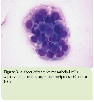
Differentiation Of Mesothelial Cells Into Macrophage Phagocytic Cells In A Patient With Clinical Sepsis

Pleural Fluid Smear Malignant Mesothelial Cells Lymphocytes Mgg Download Scientific Diagram

Figure 1 From Cytological Diagnosis Of Malignant Mesothelioma Improvement By Additional Analysis Of Hyaluronic Acid In Pleural Effusions Semantic Scholar
Pleural Fluid Mast Cells 2
Http Www Cap Org Apps Docs Committees Hematology Educational Activities 2009 Cmb Pdf
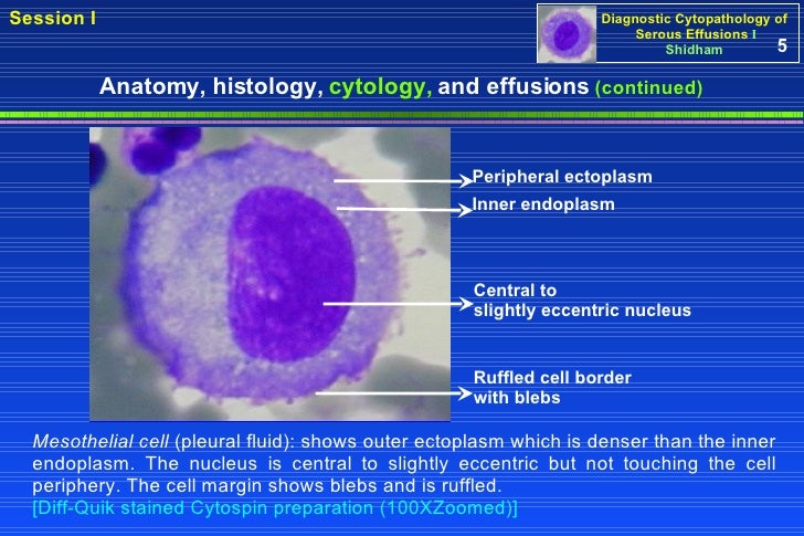
Pleural Fluid Veterian Key

The use of probrain natriuretic peptide in pleural fluid for the diagnosis of pleural effusions resulting from heart failure.
Mesothelial cells in pleural fluid diagnosis.

Effusions
Effusions Cytopathology Cellnetpathology

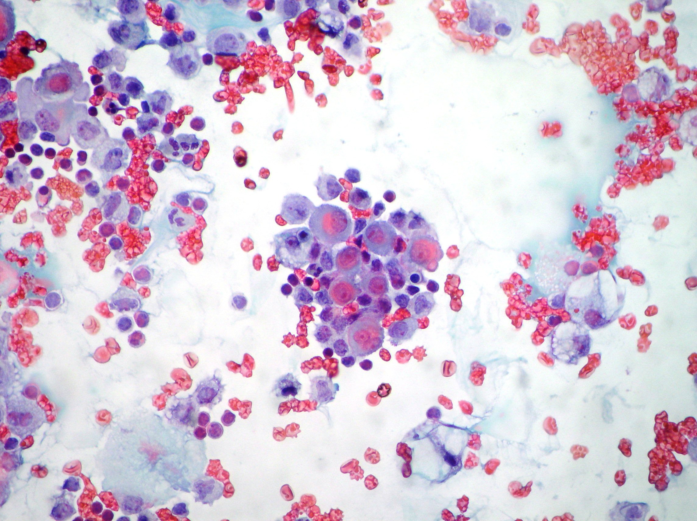
Cytology Of Pleural Fluid Clumps Of Neoplastic Cells With Download Scientific Diagram

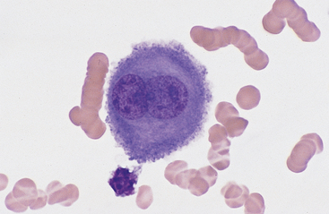
Benign Mesothelial Cells In Pleural Fluid Medical Laboratory Hematology Mad Scientist
Cytospin Processed Smear Of Pleural Fluid Revealing Uniformly Dispersed Download Scientific Diagram

The Patient S Pleural Fluid Cytology Specimen Showing A Download Scientific Diagram

A Pleural Effusion At The Time Of Diagnosis Showing Large Clusters Of Download Scientific Diagram

Pleural Fluid All Cell Blocks A D Pleural Mesothelioma Epithelial Download Scientific Diagram

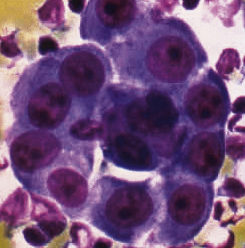
Pleural Effusion Showing Reactive Mesothelial Cells Mixed With Download Scientific Diagram

Mesothelial Cell Pleural Fluid Healthcare Medical Stock Image 653682364
Malignant And Borderline Mesothelial Tumors Of The Pleura Thoracic Key

Cytology Of Pleural And Peritoneal Lesions Chapter 5 Practical Pathology Of Serous Membranes

Diagnostic Utility Of The Cell Block Method Versus The Conventional Smear Study In Pleural Fluid Cytology

Pleural Fluid Serial Analysis Reveals Lymphocytic Predominance Few Download Scientific Diagram
Hjcam Iatrikh Zwwn Syntrofias Hellenic Journal Of Companion Animal Medicine Volume 6 Issue 1 2017 Pleural Effusion In The Cat A Focus On Laboratory Diagnosis

Mesothelial Cell Pleural Fluid Stock Photo Edit Now 652971211

Claudin 4 Expression In Mesothelial Cells From Inflammatory Pleural Download Scientific Diagram
Benign Effusions Springerlink
Https Encrypted Tbn0 Gstatic Com Images Q Tbn 3aand9gctjxi34atmipfik6awy9fwku9hnngvwavizoylai85qzyd 85bh Usqp Cau

Malignant Mesothelioma Cytology
Pathology Glossary Pleural Effusions Draw It To Know It

Results Of Pleural Fluid Diagnostics And Ct Findings Download Table

A And B Pleural Effusion Cytology A Dispersed Lymphoblasts Admixed Download Scientific Diagram
Http Www Api Pt Com Reference Commentary 2015ascope Pdf
Pleural Fluid Cytology Lab Test Dogs Vetlexicon Canis From Vetstream Definitive Veterinary Intelligence

Body Cavityfluids Chapter 3 Differential Diagnosis In Cytopathology

Figure 1 From Telomere Repeat Amplification Protocol Trap In Situ Reveals Telomerase Activity In Three Cell Types In Effusions Malignant Cells Proliferative Mesothelial Cells And Lymphocytes Semantic Scholar
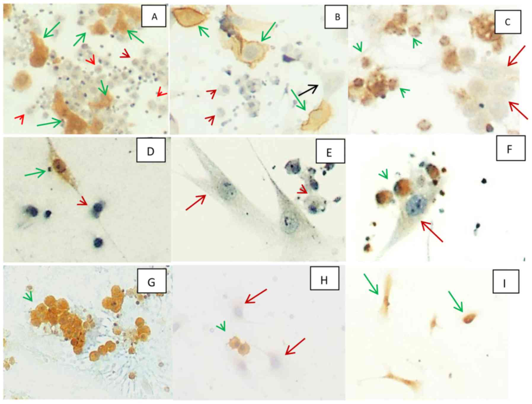
A Panel Of Markers For Identification Of Malignant And Non Malignant Cells In Culture From Effusions

Mesothelial Cell In Pleural Fluid Stock Photo Picture And Royalty Free Image Image 65541217

Mesothelial Cell In Pleural Fluid Stock Photo Picture And Royalty Free Image Image 65541187

Microfilaria Of W Bancrofti Along With Reactive Mesothelial Cells In Download Scientific Diagram
Http Www Asl5 Liguria It Portals 0 Anatomiapatologica2015 20150924 Effusion Cytology Pdf

Example Of A P16 Deletion Positive Malignant Pleural Mesothelioma Download Scientific Diagram
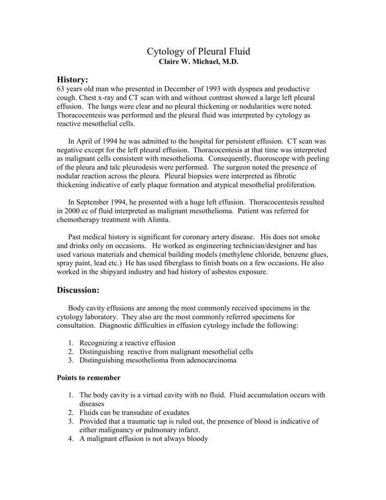
Body Cavity Effusions Are Among The Most Commonly Received

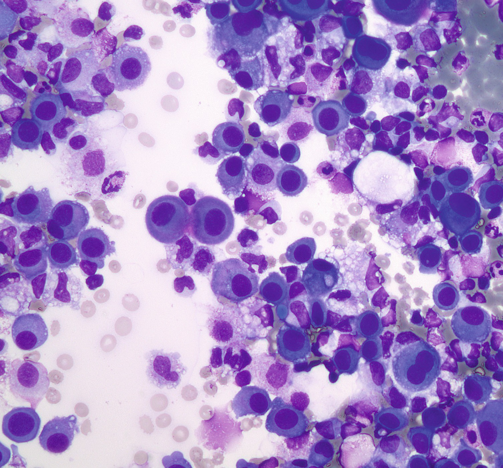
Unsuspected Multiples Myeloma Presenting As Bilateral Pleural Effusion A Cytological Diagnosis Abstract Europe Pmc
Plos One Microfluidic Purification And Concentration Of Malignant Pleural Effusions For Improved Molecular And Cytomorphological Diagnostics

Mesothelial Cell Pleural Fluid Healthcare Medical Stock Image 652849339

Mesothelial Cell Pleural Fluid Healthcare Medical Stock Image 653682337
Pleural Or Peritoneal Malignant Mesothelioma

Mesothelial Cell Pleural Fluid Stock Photo Edit Now 652971238

Mesothelial Cell Pleural Fluid Stock Photo Edit Now 652971253
Diagnostic Value Of Pleural Effusion
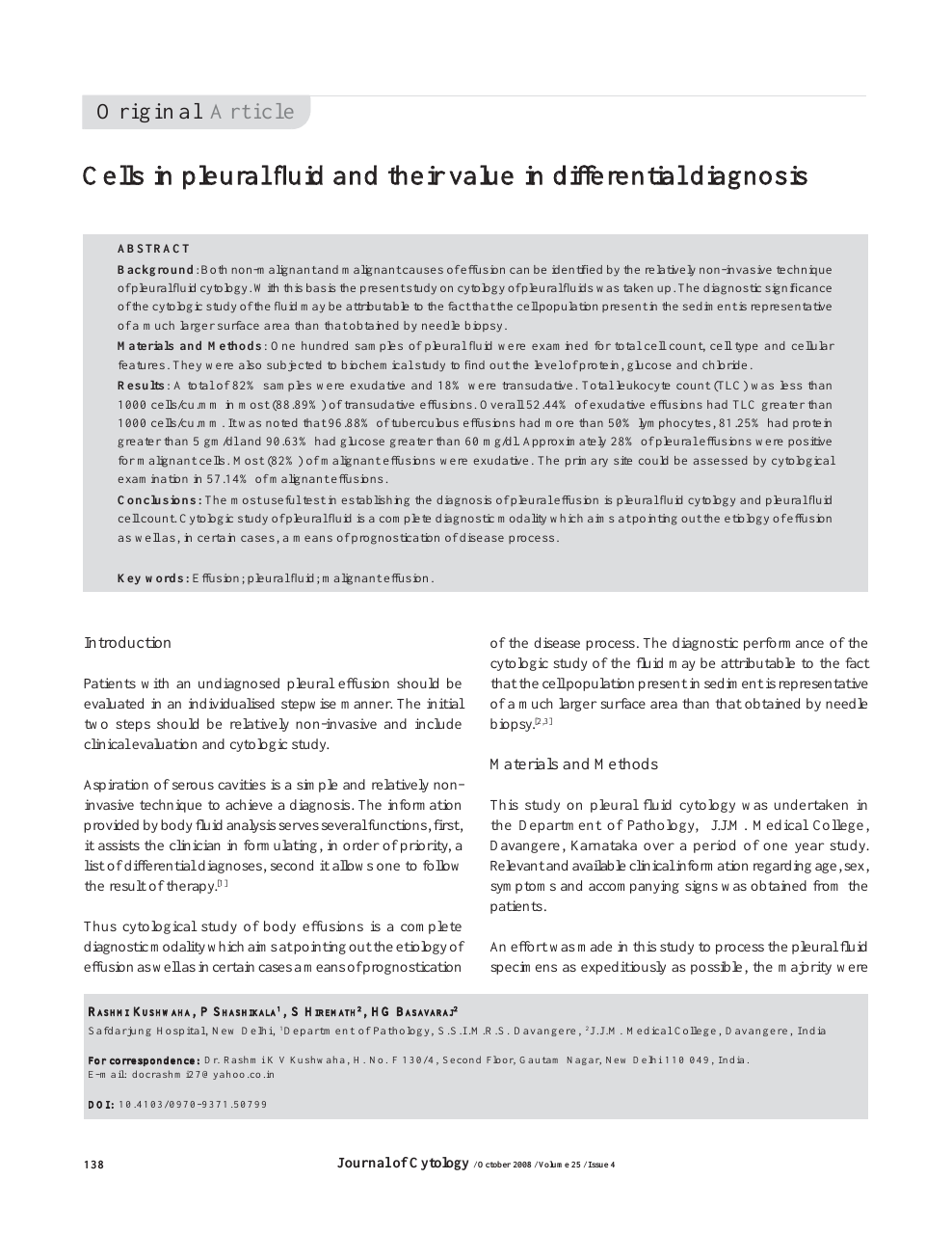
Cells In Pleural Fluid And Their Value In Differential Diagnosis Topic Of Research Paper In Clinical Medicine Download Scholarly Article Pdf And Read For Free On Cyberleninka Open Science Hub
Https Encrypted Tbn0 Gstatic Com Images Q Tbn 3aand9gcrnyd5cqson0kon9jzqmnlwqm66wbfhis Ttq6aoyy37bixsl H Usqp Cau
Source : pinterest.com
